Health By Design
 Lewis Cantley, Ph.D., and Gina DeNicola, Ph.D.
The triple-negative form of breast cancer is one of the disease's most challenging types, with lower survival rates and a more aggressive course. Pioneering cancer researcher Dr. Lewis Cantley '75, has been studying it from various angles for decades — and now, thanks to Ithaca-based engineers, he has a powerful new way to explore it from New York. "Increasingly, biological science is relying on high levels of technology to improve our ability to visualize what's going on in a tumor, and to be able to physically manipulate events within it and follow in real time what happens," says Dr. Cantley, a professor of cancer biology in medicine and the Meyer Director of the Sandra and Edward Meyer Cancer Center, who has been working with an Upstate colleague, Dr. Claudia Fischbach-Teschl, an associate professor of bio-medical engineering. "The technologies that have been developed in Ithaca are cutting-edge techniques in growing cells and even tumors in a controlled environment, where you can monitor and physically affect a tumor and look at its response. We're able to understand what's going on at a level that has never been possible in the past. It's a very exciting time."
Lewis Cantley, Ph.D., and Gina DeNicola, Ph.D.
The triple-negative form of breast cancer is one of the disease's most challenging types, with lower survival rates and a more aggressive course. Pioneering cancer researcher Dr. Lewis Cantley '75, has been studying it from various angles for decades — and now, thanks to Ithaca-based engineers, he has a powerful new way to explore it from New York. "Increasingly, biological science is relying on high levels of technology to improve our ability to visualize what's going on in a tumor, and to be able to physically manipulate events within it and follow in real time what happens," says Dr. Cantley, a professor of cancer biology in medicine and the Meyer Director of the Sandra and Edward Meyer Cancer Center, who has been working with an Upstate colleague, Dr. Claudia Fischbach-Teschl, an associate professor of bio-medical engineering. "The technologies that have been developed in Ithaca are cutting-edge techniques in growing cells and even tumors in a controlled environment, where you can monitor and physically affect a tumor and look at its response. We're able to understand what's going on at a level that has never been possible in the past. It's a very exciting time."
The joint effort between Dr. Cantley — himself a doctoral alumnus of the Ithaca campus — and Dr. Fischbach-Teschl is just one of the many ongoing collaborations between Weill Cornell Medicine researchers and Cornell engineering faculty. Research teams are working together on topics from Alzheimer's to epilepsy, reconstructive surgery to heart disease. "From my perspective, it's absolutely crucial to have collaborations with engineers," says Dr. Jason Spector, a Weill Cornell Medicine professor of surgery. "Even though I have a background in some other disciplines, at heart I'm a clinical physician and my expertise is in taking care of patients. I may have a clinical insight but no way to pursue it because I don't have the technical knowledge in that particular field. The engineers have incredible expertise in diverse areas ranging from polymer chemistry to tissue engineering to the use of lasers and optical imagery, so their insights and perspectives are perfectly complementary to mine."
Over the past decade, the university has strengthened its commitment to fostering collaborations between the two campuses, including logistical support like the Cornell bus that runs between Ithaca and New York City several times a day, providing reliable Wi-Fi, snacks and a comfortable place to work during the 200-mile journey. Occasional retreats between engineers and their counterparts in Weill Cornell Medicine departments like surgery, neurological surgery, and radiology allow faculty to make matches with colleagues who have complementary research interests. The university has also offered several rounds of seed grant funding to help intercampus projects get off the ground — and according to one initial analysis, the investment has more than paid off.
In 2010, Dr. Lawrence Bonassar, a professor of biomedical engineering and ofmechanical and aerospace engineering, studied the outside funding generated by roughly a dozen awards given in the previous five years, finding that the seed support had facilitated substantive results that impressed outside granting agencies. "It had about an eight-fold return on investment," reports Dr. Bonassar, whose collaborative projects with Weill Cornell Medicine surgeons include two types of implants made of living tissue: ears for children born without them, and replacements for degenerated spinal discs that cushion the vertebrae. "About $400,000 in grants had been given out — and they had returned, at that time, more than $3 million from other organizations including the National Institutes of Health, private foundations, and the National Science Foundation. So these tend to be productive collaborations that are well received by the outside world."
 Dr Evi Giannakakou in her lab
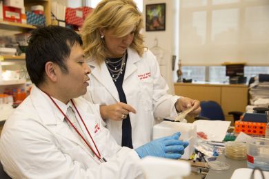
In the field of oncology research, intercampus collaborations got a particularly strong boost in 2009 with the awarding of a five-year, $13 million grant from the National Cancer Institute that created the Center on the Microenvironment and Metastasis. Established under the NCI's Physical Sciences-Oncology Centers initiative, it has fostered numerous projects including the ongoing effort by Weill Cornell Medicine's Dr. David Nanus, the Mark W. Pasmantier Professor of Hematology and Oncology in Medicine, and Dr. Evi Giannakakou, an associate professor of pharmacology, to develop a method to detect prostate cancer cells in the blood. They're working with Dr. Brian Kirby, an associate professor of mechanical and aerospace engineering who is based in Ithaca but holds a joint appointment in Weill Cornell Medicine's Division of Hematology and Medical Oncology. Says Dr. Kirby: "There's basically no way for me to impact human health directly unless I'm working with clinicians, looking at real samples, and trying to influence actual clinical practice."
Dr Evi Giannakakou in her lab
In the field of oncology research, intercampus collaborations got a particularly strong boost in 2009 with the awarding of a five-year, $13 million grant from the National Cancer Institute that created the Center on the Microenvironment and Metastasis. Established under the NCI's Physical Sciences-Oncology Centers initiative, it has fostered numerous projects including the ongoing effort by Weill Cornell Medicine's Dr. David Nanus, the Mark W. Pasmantier Professor of Hematology and Oncology in Medicine, and Dr. Evi Giannakakou, an associate professor of pharmacology, to develop a method to detect prostate cancer cells in the blood. They're working with Dr. Brian Kirby, an associate professor of mechanical and aerospace engineering who is based in Ithaca but holds a joint appointment in Weill Cornell Medicine's Division of Hematology and Medical Oncology. Says Dr. Kirby: "There's basically no way for me to impact human health directly unless I'm working with clinicians, looking at real samples, and trying to influence actual clinical practice."
Last summer's establishment of the Meinig School of Biomedical Engineering also strengthened intercampus research — elevating an existing department that had been founded in 2004. The school was created with a $50 million gift from undergraduate alumni Nancy and Peter Meinig and their children; as Weill Cornell Medicine Dean Dr. Laurie Glimcher, said at the time, their generosity "will enable us to expand our collaborations and advance our critical work translating new discoveries into the best patient care."
Weill Cornell Medicine researchers are also working with Cornell engineers who are based in Manhattan. Not only does the new Cornell Tech campus (temporarily located in Chelsea, but with three buildings currently under construction on Roosevelt Island) have a program devoted to health-related topics, but about 10 percent of students earning doctorates in biomedical engineering at the university are doing their thesis research at Weill Cornell Medicine. "When you look around the country, you'll see that there are no biomedical engineering programs in the top 10 or 20 that aren't strongly tied to a top medical school — and interestingly, there aren't many top medical schools that aren't strongly tied to a bio-medical engineering program," says Dr. Chris Schaffer, an Ithaca-based associate professor and director of graduate studies in biomedical engineering, noting that all of his department's first-year doctoral candidates spend about two months at Weill Cornell Medicine for an immersion program in clinical medicine. "Much of the future of biomedical research, and of improving healthcare, will involve increased reliance on quantitative and engineering approaches to drugs and diagnostics. We do that best when we do it together."
EARS FOR KIDS
Of every 10,000 babies born worldwide, between one and four have a congenital birth defect called microtia, in which one or both outer ears is missing or deformed. Unfortunately for these kids and their families, the options for surgical reconstruction have long been limited and imperfect — consisting of either an artificial implant or one crafted from the patient's rib cartilage. But professor of surgery Dr. Jason Spector, and biomedical engineer Dr. Lawrence Bonassar, hope they'll soon be able to offer an alternative: a natural implant created from a patient's own cells that would grow along with the child and never require replacement. For several years, the two have been working together on the ears, which could either be formed through 3D printing or shaped in a mold. Since most cases of microtia occur on only one side of the head, computer imaging can be used to scan a patient's normal ear, so the implants can be custom-made for a perfect fit. While the work began using bovine tissue, it has evolved to comprise human cartilage; microtia patients usually have a small amount of vestigial ear tissue, which can be nurtured with stem cells until there is enough material to form an implant. The results have been highly promising: the growth system is working well, and the resulting ears are durable and have the proper anatomical form. The researchers hope to move into clinical trials in the next two to three years. "As a result of our collaborative efforts, we are poised to revolutionize the treatment of children who have microtia — and as we further develop these cutting-edge tissue engineering strategies, to make much needed 'replacement parts' in many other areas of the body," Dr. Spector says. "It's all very exciting."
TUMOR-CELL DETECTOR
It's known as a "liquid biopsy": taking a sample of blood from a patient and analyzing it for cancer cells. Potential uses for such technology include allowing oncologists to refine a diagnosis, detecting the spread of cancer from a tumor to elsewhere in the body, and predicting the efficacy of a certain drug — all without an invasive procedure. According to Dr. David Nanus, associate director of clinical services at the Meyer Cancer Center, "it's something that a lot of medical centers and cancer centers are working on that will have utility in the future." Dr. Nanus has been working with pharmacologist Dr. Evi Giannakakou, and engineer Dr. Brian Kirby, on employing liquid biopsies for a particular purpose: they're doing a proof-of-concept study using a microfluidic device to detect prostate cancer circulating tumor cells. An expert in microfluidics, Dr. Kirby devised a silicon chip known as GEDI; pronounced like the Star Wars heroes, it stands for Geometrically Enhanced Differential Immunocapture. "We coat the surface of the microdevice with a substance that will specifically stick to cancer cells and not to blood cells," Dr. Kirby explains. "We designed it to be sort of a maze that tricks the cancer cells into colliding with the surfaces many times over." The multi-institution study, involving about five dozen patients with late-stage prostate cancer, is looking for biomarkers that may predict sensitivity or resistance to a class of chemotherapy drugs known as taxanes. "A major goal is to figure out the mechanism of chemotherapy resistance," Dr. Nanus says. "If a patient progresses, can we look at their circulating tumor cells and — from an analysis of their DNA or RNA expression — determine the molecular reason why they have become resistant to treatment?"
BATTLING BREAST CANCER
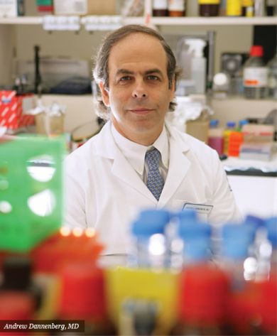
The "seed and soil" concept of cancer metastasis holds that tumors require fertile ground in which to grow — which means that making the body inhospitable to them is key to battling the disease. Dr. Andrew Dannenberg, the Henry R. Erle, M.D.- Roberts Family Professor of Medicine, and breast cancer specialist Dr. Linda Vahdat, have ongoing collaborations with biomedical engineer Dr. Claudia Fischbach-Teschl, whose Ithaca lab is devoted to studying the environment in which a tumor exists, including its surrounding blood vessels and other non-tumorous host cells. They have worked together to explore diagnosis, treatment and prevention from two major angles: the effect of obesity on patient outcomes and — a major focus of Dr. Vahdat's work — the promise of copper-depleting drugs in treating triple-negative breast cancer. For the obesity work, for example, Dr. Vahdat provided specimens from lean and obese breast cancer patients; Dr. Fischbach-Teschl was able to show that obese women have increased levels of proteins typically associated with wound-healing, which makes for a "soil" more hospitable to cancer, thus raising the likelihood of its spread. "Claudia's expertise is in physical factors that promote metastasis — getting down to the practical aspects of, 'How does a tumor cell get from point A to point B?' says Dr. Vahdat, a professor of medicine. "It's a totally different way to look at things, but it's highly complementary to what someone like myself does, which is on a very macro level, taking care of patients."
BACK PAIN RELIEF
Among the projects sparked during a retreat between Ithaca engineers and Weill Cornell Medicine surgeons is one that could someday provide relief to the millions of Americans suffering from chronic back or neck pain. Spinal surgeon Dr. Roger Härtl, is working with Dr. Bonassar to develop a bioengineered intervertebral disc to replace those lost to degeneration or herniation — be it from natural aging, playing contact sports, or other factors like obesity or years of manual labor. Currently, most patients are treated with pain medication or physical therapy — but 5 to 10 percent require more invasive methods such as injections or surgery. In the latter case, the most common procedure is spinal fusion, in which a damaged disc is removed and bone is fused into the gap; while artificial discs made of metal and plastic are available, they can damage adjacent tissue, and their longevity is unclear. So Dr. Härtl, a professor of neurological surgery and director of spinal surgery and neurotrauma at Weill Cornell Medicine's Brain and Spine Center, teamed up with Dr. Bonassar to design 3D-printed implants made of living tissue. The technology was successfully demonstrated in rats; in collaboration with the College of Veterinary Medicine, it's currently being tested in animals, who can suffer from disk degeneration just as humans do. Ultimately, the implants could be created based on MRI imaging of a patient's spine and comprise cells either taken from a cadaver or — a greater challenge — grown using a person's own tissue. Another project, which Dr. Härtl reports is much closer to clinical implementation, is a technique to repair injured discs using tissue-engineered biological "glues."
A 'LASER SCALPEL' FOR EPILEPSY
Of the two main kinds of epilepsy — general and focal — the latter is much harder to treat. While general epilepsy — in which the entire brain spasms with neural activity — can be well controlled with medication, focal epilepsy is more challenging. Often caused by brain trauma or congenital malformation, it involves too much neural activity in specific regions; to control it with drugs, says biomedical engineer Dr. Chris Schaffer, "you end up almost anesthetizing the brain." Instead, the condition is treated with resective brain surgery — an imprecise option that can lead to cognitive and motor deficits. But Dr. Schaffer, an expert in advanced optical techniques, is working with Dr. Theodore Schwartz, the David and Ursel Barnes Professor of Minimally Invasive Neurosurgery, to offer a much better surgical solution. They're collaborating to develop a "laser scalpel" that can cut with precision on the scale of a micron — one-millionth of a meter. They're writing up a paper reporting promising results in a rodent model, in which a targeted cut decreased seizures by 50 percent; they're currently studying whether the improvement is long-lasting. Dr. Schaffer is also working with Dr. Frank Wise '88, the Samuel B. Eckert Professor of Engineering in Ithaca's Department of Applied and Engineering Physics, to design the device, which pulses at a rate of 100 femtoseconds. "The ratio of 100 femtoseconds to a minute is about the same as the ratio of a minute to the known age of the universe [about 13.8 billion years]," Dr. Schaffer notes. "So it's a really, really short burst of light."
HEART OF THE MATTER
At the Dalio Institute of Cardiovascular Imaging, director Dr. James Min, and colleagues aim to better understand heart disease, in the hope that it will lead to improved treatments and preventive measures. To that end, they use not only familiar tools like MRI, CT and PET — but also novel technologies like 3D printing and computer modeling of blood flow dynamics, drawing on the expertise of Ithaca-based engineers. Dr. Min, a professor of radiology and of medicine who is board certified in cardiology, has been working with Dr. Jonathan Butcher, associate professor of biomedical engineering and associate director of undergraduate studies in biomedical engineering, to study how the particulars of various surgical procedures — say, the shape and placement of a vascular graft — affect patient outcomes. Ultimately, grafts that have been designed for optimum performance in an individual person's heart could be fabricated through tissue engineering, obviating the need to obtain them from elsewhere in a patient's body. The grafts — or even entire heart valves — would be made of a patient's own cardiac cells, harvested by surgeons and grown on a scaffold of water-soluble polymers. In the best-case scenario, Dr. Butcher says, "we could do all of those procedures in the operating room, in one sitting." On the clinical side, the collaboration also involves veteran heart surgeon Dr. Leonard Girardi '89, chair of the Department of Cardiothoracic Surgery and cardiothoracic surgeon-in-chief at NewYork-Presbyterian/Weill Cornell Medical Center.
This story first appeared in Weill Cornell Medicine, Vol. 15, No.1.