Engineering tumor environments for study
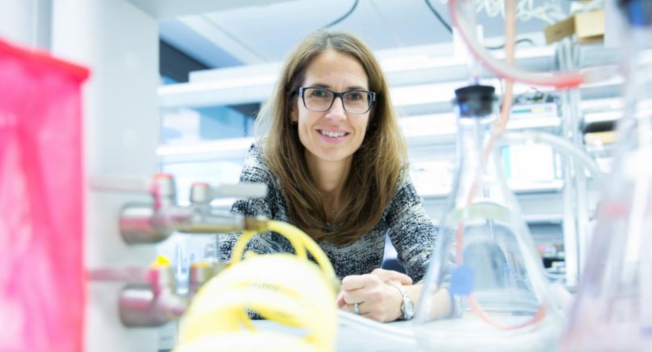
 Claudia Fischbach-Teschl, Ph.D.
A cancer tumor does not grow on a petri dish, so to understand how tumor cells function, it’s important to study tumors under conditions in which they naturally exist.
Claudia Fischbach-Teschl, Ph.D.
A cancer tumor does not grow on a petri dish, so to understand how tumor cells function, it’s important to study tumors under conditions in which they naturally exist.
How do tumors change normal stem cell functions in the body? How do breast cancer cells exploit bone cells to develop bone metastasis? What type of vasculature do tumor cells need to spread throughout the body? Claudia Fischbach-Teschl, Biomedical Engineering, is working to answer such questions, using tissue-engineered model systems that elucidate the effect of the tumor cell’s natural microenvironment in these situations.
Tumors' Microenvironment
The microenvironment is essentially the neighboring tissue and its reconstituting components that surround our body’s cells. Cells don’t exist individually but are surrounded by other cells and the extracellular matrix, a bioactive material that not only functions as a biological glue to keep cells together but also regulates cell signaling.
“We’re interested in how cells interact within this matrix and which physical as well as biochemical cues may regulate these interactions,” Fischbach-Teschl says.
“You can plate cells on a plastic surface (as in petri dishes) and study their behavior, but this approach lacks tissue context and thus, may not fully recapitulate the in vivo situation.”
Fischbach-Teschl’s lab has developed 3-D scaffolds and model systems to provide cells with this environment in order to study cancer under physiologically more relevant conditions.
How Breast Cancer Cells Exploit Bone Properties
In one research project, Fischbach-Teschl’s lab uses polymer scaffolds that include minerals to mimic certain aspects of the bone's extracellular matrix. In collaboration with Lara A. Estroff, Materials Science and Engineering, the researchers made sponge-like scaffolds that contain the minerals to mimic different bone makeups. They then use the scaffolds to study the effect of varying bone mineral properties on breast cancer cell localization to bone.
This is important because most people do not die from primary cancer but from tumors that spread throughout the body, especially to the bone. Eighty percent of breast cancer patients develop bone metastasis, Fischbach-Teschl says. The goal is, therefore, to figure out how breast cancer cells exploit bone material properties and cell functions and which molecules are involved in these processes.
Mechanical stimulation intrinsic to bone also plays a key role in how cancer cells propagate. In collaboration with Marjolein van der Meulen, Mechanical and Aerospace Engineering/Biomedical Engineering, and Lawrence Bonassar, Biomedical Engineering, Fischbach-Teschl's lab is looking at how mechanical loading, such as walking and exercise, affects the progression of bone metastasis.
The researchers have found compelling evidence that weight-bearing exercise could have anti-metastatic effects in bone. They injected tumor cells into bones of mouse models, which were then mechanically stimulated. They also injected tumor cells into porous scaffolds and then squeezed the scaffolds to mimic compressive mechanical loading, which occurs during exercise. Under these circumstances, they found that mechanical loading inhibited a gene, Runx2, involved in cancer, and this may be the reason for reduced metastasis. Fischbach-Teschl’s next goal is to test this in patients.
How Tumors Affect the Microenvironment of Normal Stem Cells
Fischbach-Teschl also wants to understand how tumors modulate the microenvironment in which normal stem cells reside. Typically, stem cells contribute to physiological functions, such as tissue development, wound healing, and regeneration. Tumors may alter the properties of these cells, however, in a manner that ultimately worsens the cancer.
In one study the researchers found that in the presence of tumors, adipose-derived stem cells–those in fatty tissue–are more susceptible to becoming myofibroblasts, cells that mediate the typical stiffening of tumors, enabling their detection via palpation. This is particularly important in breast cancer patients who want reconstructive surgery after a mastectomy. Doctors often use adipose tissue to reconstruct breast tissue.
Fischbach-Teschl and her lab, however, found that if the contained adipose-derived stem cells come in contact with any residual cancer cells, they could promote recurrence of cancer instead of developing into new, healthy breast tissue.
Developing Cancer Models to Answer Scientific Questions
In all of her research, Fischbach-Teschl says that she is motivated to develop models to answer specific scientific questions. “I’m excited about the basic science questions that can be answered with these models and that may eventually change therapy,” she says. “Initially, we developed cancer models that were very simple–just tumor cells seeded into a three-dimensional polymer scaffold rather than a petri dish–and we saw dramatic differences in cell behavior. That provided the basis for us to build more complex models.”
It is evident that Fischbach-Teschl’s work has clear clinical relevance. She says that in the future, she hopes to make models that provide a more realistic tissue context for testing patient-specific drug therapy–for example, a model system could be seeded with a patient’s cells and accurately show how that patient will respond to a specific treatment without having to take the drug. She also hopes to develop more complex systems that integrate blood vessels in order to study transport phenomena in tumors that compromise effective drug therapy.
Fischbach-Teschl stresses the importance of strong collaborations with her colleagues at Cornell in Ithaca and Weill Cornell Medicine. “We can do great engineering, but to create something that may ultimately change a patient's life,” she says, “It’s important for us to have clinical connections and insights.”
This feature first appeared on the Cornell Research site.